iCare MAIA TrueColor Microperimetry
iCare

iCare MAIA TrueColor Microperimetry

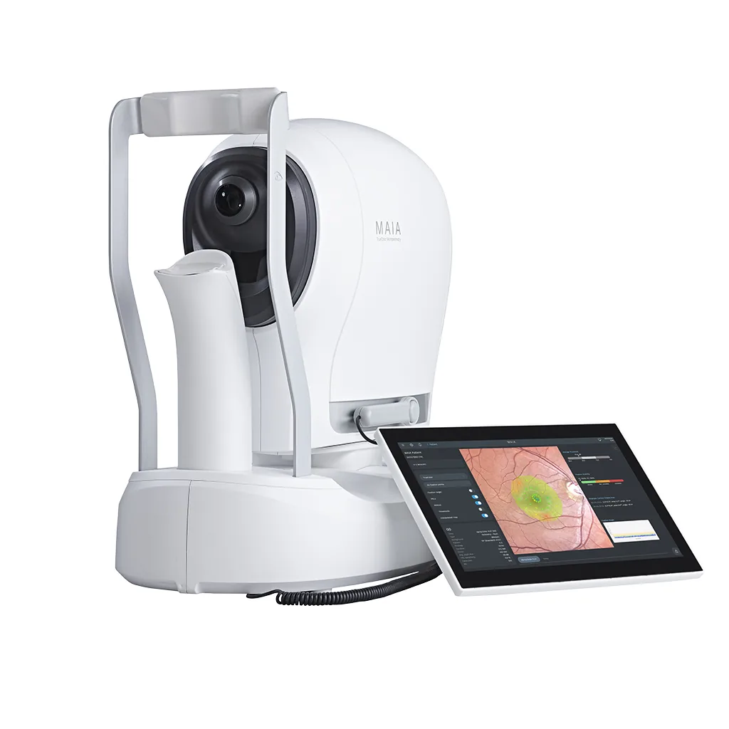
OCT tells you what a macula looks like. The MAIA tells you what it can still do. In one automated exam it maps retinal sensitivity point by point, tracks the fundus at 25 Hz so each point lands where intended, and captures a non-mydriatic confocal image of the same retina: function and structure from a single sitting. The imaging side spans TrueColor, infrared, red-free, blue and red modalities across a 60-degree field, and the test grids are customizable to the pathology in front of you. For AMD and macular disease practices, that pairing does two jobs: it sharpens diagnosis and monitoring, and it gives patients a picture of their own function worth staying in treatment for.

Request Pricing → Schedule a Demo →
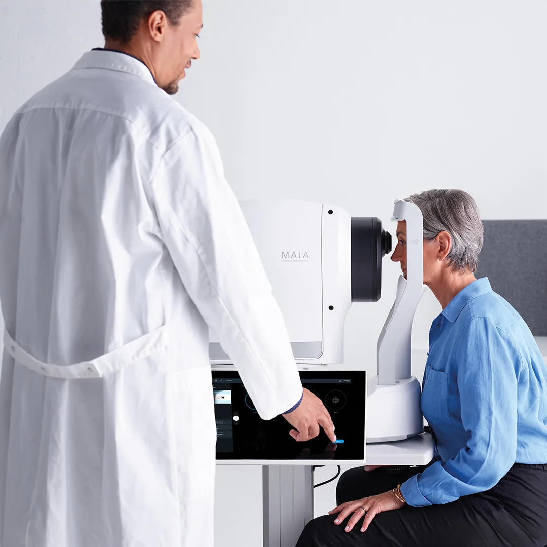
Doctor performing iCare MAIA microperimetry exam with patient
Practice Fit

Where The MAIA Wins

The MAIA belongs in practices where connecting structure to function, and showing patients both, changes how macular disease gets managed.

Best Fit

Retina and macular disease practices managing AMD, diabetic macular edema and related pathology, especially where keeping patients engaged in long-course therapy is half the clinical battle.

Why This

A patient can have a dramatic OCT and functional vision, or a quiet OCT and real functional loss. Structure alone does not arbitrate. Microperimetry does, point by point, and the MAIA’s real-time tracking keeps the mapping accurate even when advanced disease has destabilized fixation.

Practice Impact

Injection fatigue is real, and “the OCT looks stable” is a hard sentence to stay in treatment for. A sensitivity map of the patient’s own macula, holding steady because the therapy is working, makes the case in a way patients can see.

Consider Instead

The MAIA measures macular function over a small central field. If you need standard 24-2 or 30-2 threshold perimetry for glaucoma, that is the COMPASS or the VF2000, not this instrument. Some retina practices run the MAIA alongside a perimeter rather than instead of one.

Platform Strengths

Why Practices Choose MAIA

Structure Meets Function

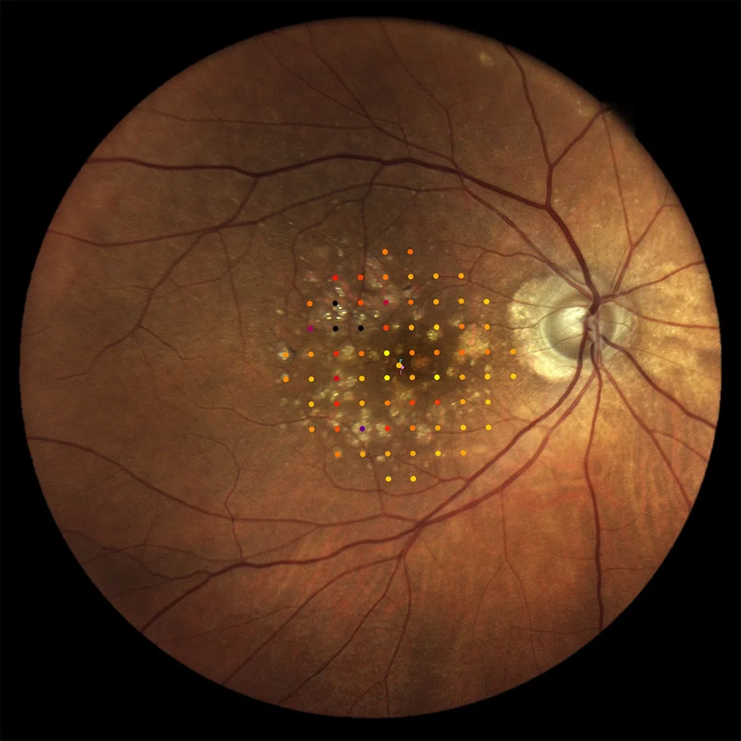
The sensitivity map sits on the TrueColor image of the patient’s own fundus: where the disease is and what it costs them, in one picture.

Real-Time Retinal Tracking

Advanced macular disease destabilizes fixation, exactly when accurate mapping matters most. The 25 Hz tracker compensates in real time, so those patients still produce usable data.

Reduce Patient Drop-Off

Progression reports over time turn “keep coming in” into a chart the patient can read: this is your function, and this is what the treatment is preserving.

What comes out of the exam

Sensitivity, fixation, and the reports they land in

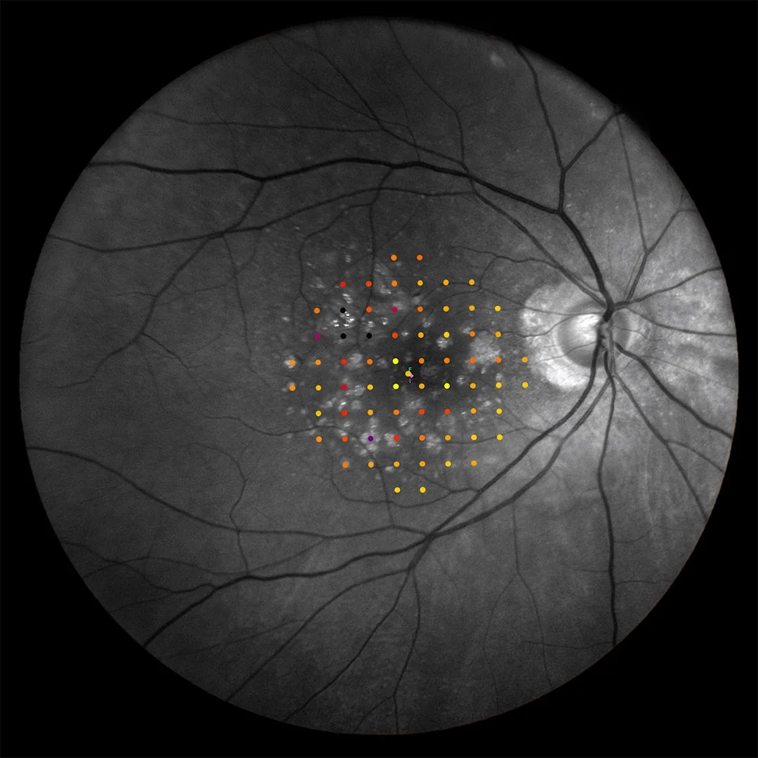
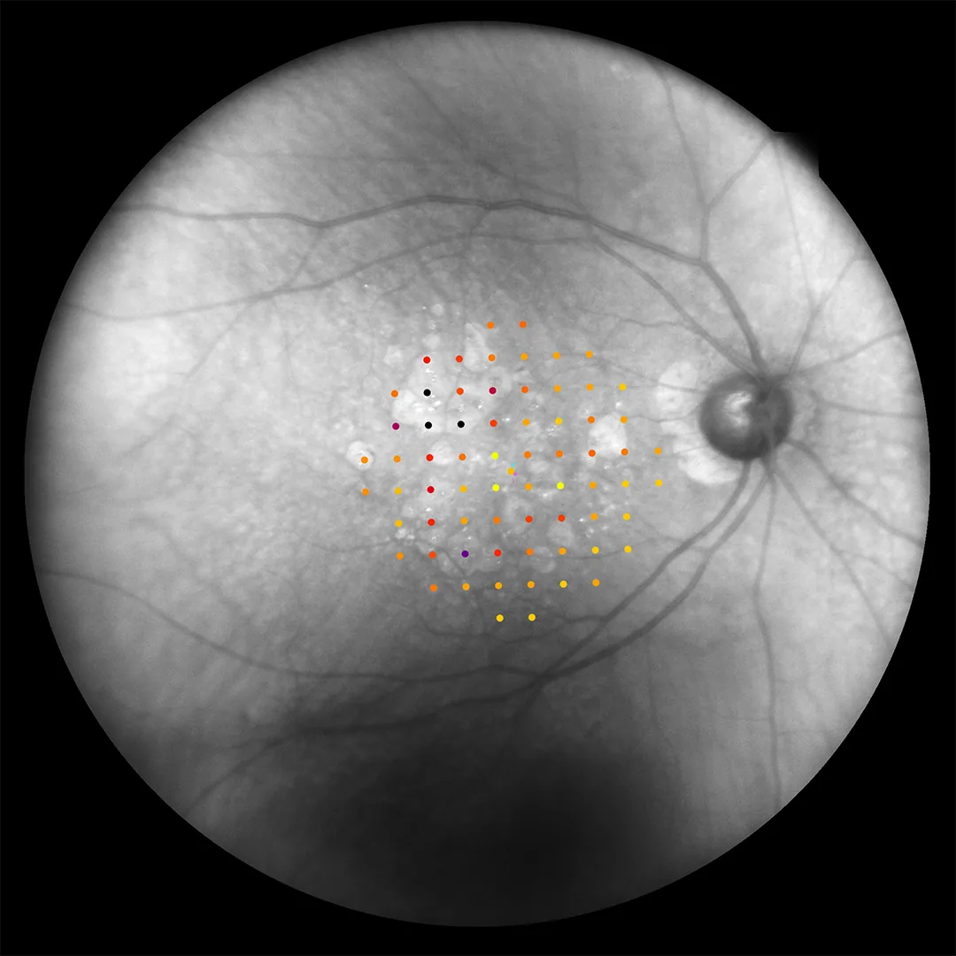
The same macula rendered three ways with the sensitivity grid laid over each: TrueColor, infrared, and red-free.

iCare MAIA TrueColor microperimetry grid overlay showing retinal sensitivity

TrueColor Grid Overlay: Sensitivity map overlaid on TrueColor fundus image

iCare MAIA red-free microperimetry grid showing vascular detail

Red-Free Grid: Sensitivity mapping with enhanced vascular visualization

iCare MAIA infrared microperimetry grid for sub-surface detail

Infrared Grid: Sensitivity data with sub-surface retinal detail

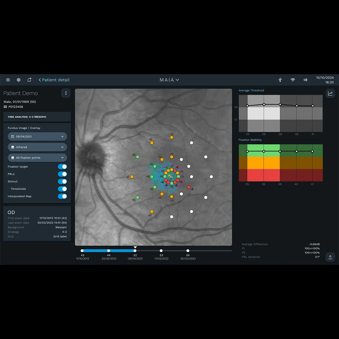
iCare MAIA time analysis showing mesopic sensitivity progression

Time Analysis: Mesopic sensitivity progression tracking over time

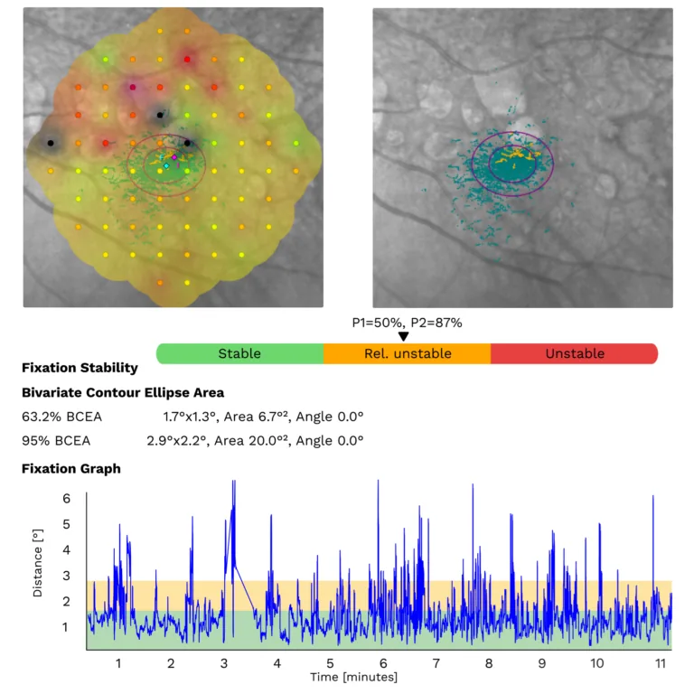
iCare MAIA fixation stability analysis showing preferred retinal locus

Fixation Analysis: Fixation stability and preferred retinal locus mapping

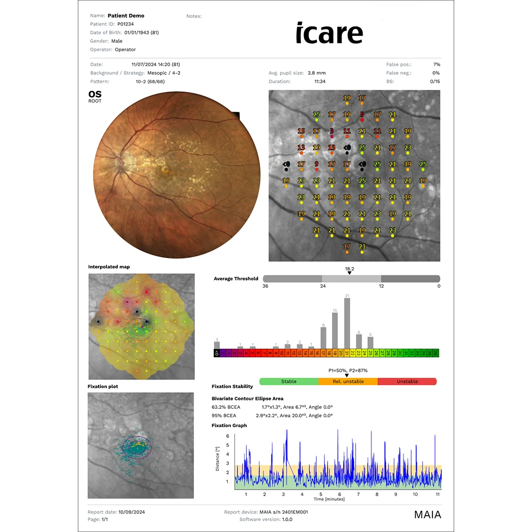
iCare MAIA examination report with sensitivity and fixation data

Examination Report: Comprehensive sensitivity and fixation reporting

Clinical images and examination reports courtesy of iCare / Revenio Group.

Specifications

Technical Details

Technology
TrueColor confocal microperimetry
Projection Field
30°
Background Luminance
4 asb
Maximum Luminance
1000 asb
Dynamic Range
36 dB
Stimulus Size
Goldmann III
Stimulus Duration
200 ms
Retinal Tracking
25 Hz automated
Test Strategies
Full threshold (4-2), Suprathreshold, Fixation only
Test Grids
10° (37 pts), 6° (37 pts), 20° (41 pts), 20° 10-2 pattern (68 pts), Custom
Fundus FOV
60°
Sensor Resolution
5 MP (2592 × 1944)
Imaging Modalities
TrueColor, Infrared, Red-free, Blue, Red
Optical Resolution
17 µm
Min. Pupil Size
3 mm (non-mydriatic)
Auto Refraction
–12D to +15D
Display
15" multi-touch
Connectivity
1 Gbit Ethernet, Wi-Fi (802.11ac/b/g/n), USB export
Storage
SSD 480 GB+
Dimensions (W × H × D)
360 × 590 × 620 mm (14.2" × 23.2" × 24.4")
Weight
27 kg (59 lbs)
Power
100–240 VAC, 50–60 Hz, 80W
Classification
IEC 60601-1 Class II, Type B

Specifications reproduced from the manufacturer's published materials and last checked on September 1, 2026. Configurations and specifications change; confirm against the current quote before you rely on them.

The Whitecotton Vision Advantage

What You Get Beyond the Box

Installed in Your Lanes

Delivery is scheduled around your clinic days, the device is configured in your actual exam space, and your staff practices on it hands-on before the visit ends.

Workflow Planning

Where does the device live, who runs it, and how do results reach the doctor? That capture-to-review path gets mapped with your staff before go-live, so the technology fits how your clinic already moves.

Direct Support

When you need help you reach Ryan directly. No call center, no ticket queue, no account manager you have never met.

Ready to see the MAIA in action?

Talk to Ryan for pricing and configuration options, or to see it running in your own pretest flow before you decide.

Request Pricing → Schedule a Demo →
Call Ryan Email About This