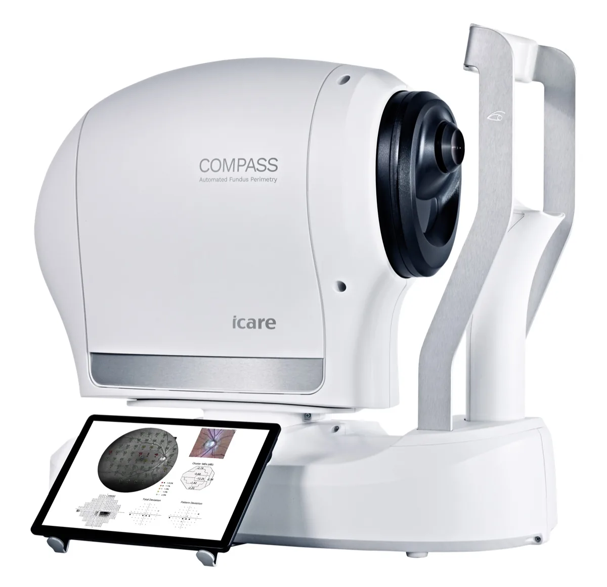
COMPASS Perimeter
iCare

COMPASS Perimeter

Traditional perimetry runs on an assumption: that the patient held fixation, so each stimulus landed where the test map says it did. COMPASS removes the assumption. A built-in confocal scanning ophthalmoscope watches the retina at 25 frames per second throughout the test, records a simultaneous 60° fundus image, and ties every threshold point to a verified retinal location. When the field and the fundus come from the same instrument in the same sitting, progression calls rest on data you can defend, which is the whole point for a glaucoma practice.

Request Pricing → Schedule a Demo →
Practice Fit

Where COMPASS Wins

COMPASS earns its place when the practice wants field results anchored to the retina that produced them.

Best Fit

Glaucoma-focused practices, advanced disease management, neuro-ophthalmic workups, and any doctor tired of explaining away unreliable fields.

Why This

Ask what your current perimeter does when the patient’s eye wanders. If the answer is “flags it afterward,” that is the difference: COMPASS compensates during the test, tracking the retina at 25 fps, and hands you a fundus image from the same sitting so structure and function overlay without a second instrument.

Practice Impact

Fewer fields get repeated for reliability, progression conversations rest on defensible data, and the glaucoma workup both feels and is more rigorous.

Consider Instead

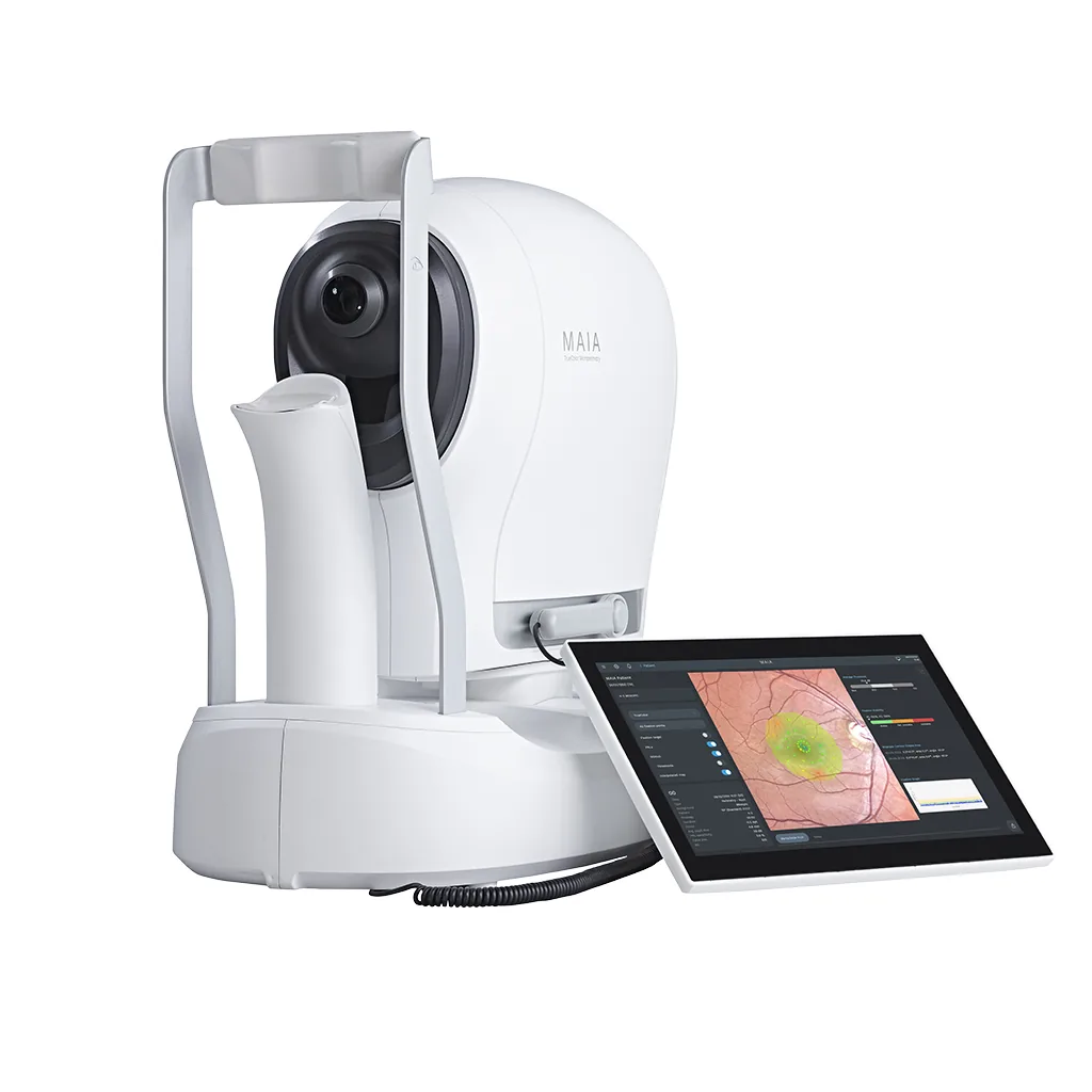
If the constraint is a room you do not have, look at the Micro Medical VF2000. A headset needs no dedicated lane and travels to satellite sites. If the question is macular function rather than a full field, the MAIA answers it more directly.

Clinical Confidence

Trust What You Measure

Retinal Tracking

The tracker runs at 25 Hz for the entire test, so each threshold point maps to where the retina actually was, not where the fixation target hoped it would be.

Structure + Function

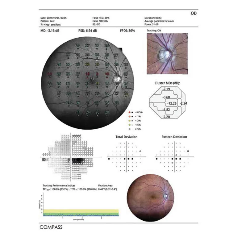
Sensitivity data lands directly on the anatomy that produced it: the confocal fundus image comes from the same test, so there is no cross-instrument alignment to trust.

Smart Progression

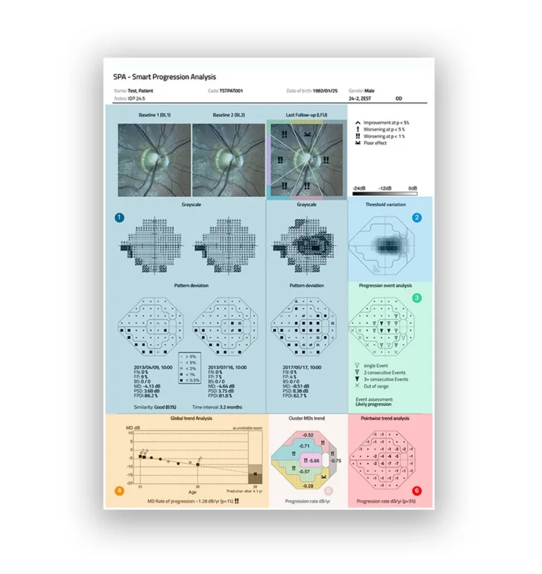
Smart Progression Analysis reads longitudinal change two ways, cluster-based and pointwise trends, which is how real progression gets separated from test-retest noise.

What the report looks like

The field and the fundus on one page

Every test produces two things at once: threshold sensitivity, and a confocal photograph of the retina those thresholds were measured on. Below are the outputs a doctor actually reads.

COMPASS Smart Progression Analysis report showing visual field trends

Smart Progression Analysis (SPA): Change over time, read two ways at once: deviation by cluster, and rate of change point by point.

COMPASS visual field report with integrated fundus image

Visual Field Report with Fundus Overlay: Sensitivity values printed onto the 60° retinal image they were measured from, so structure and function are read side by side.

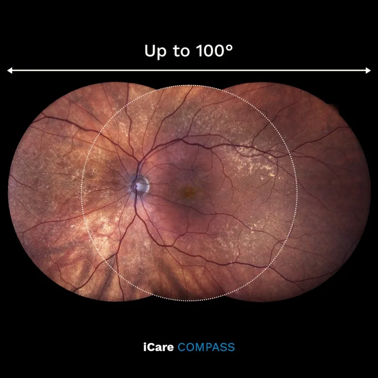
COMPASS SmartMosaic panoramic fundus image

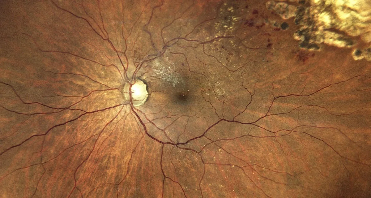
100° SmartMosaic Fundus Image: High-resolution TrueColor confocal panoramic view acquired during visual field testing. No additional imaging session required.

Report examples and clinical images courtesy of iCare / Revenio Group.

Specifications

Technical Details

Projection Field
30° radius
Dynamic Range
0–50 dB
Background Luminance
31.4 asb / Max: 10,000 asb
Stimulus
Goldmann III, round, white, 200 ms
Fixation Control
25 Hz automated retinal tracking
Imaging FOV
60° diameter (100° horizontal with SmartMosaic)
Imaging Sensor
5 megapixels (2592 × 1944) / 17 µm resolution
Light Sources
IR LED (825–870 nm) / White LED (440–650 nm)
Imaging Modalities
Color, infrared, red-free, stereoscopic, SmartMosaic
Auto-focus Range
–12D to +15D (no trial lenses)
Display
Tablet-operated multi-touch color display
Storage
SSD ≥480 GB
Dimensions (W × D × H)
360 × 620 × 590 mm
Weight
25 kg (55 lbs)
Power
100–240 VAC, 50–60 Hz, 80W / IEC 60601-1 Class I

Specifications reproduced from the manufacturer's published materials and last checked on September 1, 2026. Configurations and specifications change; confirm against the current quote before you rely on them.

The Whitecotton Vision Advantage

What You Get Beyond the Box

Installed in Your Lanes

Delivery is scheduled around your clinic days, the device is configured in your actual exam space, and your staff practices on it hands-on before the visit ends.

Workflow Planning

Where does the device live, who runs it, and how do results reach the doctor? That capture-to-review path gets mapped with your staff before go-live, so the technology fits how your clinic already moves.

Direct Support

When you need help you reach Ryan directly. No call center, no ticket queue, no account manager you have never met.

Ready to see the COMPASS in action?

Talk to Ryan for current pricing and availability, or to put one in your exam lane and try it on your own patients.

Request Pricing → Schedule a Demo →
Compare COMPASS vs HFA3 vs Octopus 900 →
Call Ryan Email About This